Zur diesjährigen MICCAI (International Conference on Medical Image Computing and Computer Assisted Intervention) vom 27.09. bis 01.10.2021 werden die Forschenden um Julian Alpers und Daniel Reimert ihre neuen Ergebnisse zur bildgeführten Tumorablation mittels 2,5D-Thermometrie-Bildgebung im MRT vorstellen. Mit einem Impact Faktor von 8,5 ist diese Konferenz eine der international wichtigsten und weitreichendsten Konferenzen im Themengebiet „Medizinische Bildverarbeitung und computergestützte Intervention“. Insgesamt widmet sich die MICCAI Society der Förderung, Erhaltung und Erleichterung von Forschung, Ausbildung und Praxis auf dem Gebiet der medizinischen Bildverarbeitung und computergestützter medizinischer Interventionen, einschließlich biomedizinischer Bildgebung und medizinischer Robotik.
Der Beitrag des Forschungscampus STIMULATE behandelt den dringenden klinischen Bedarf einer schnellen und zuverlässigen Überwachung der volumetrischen Wärmeverteilung während der MRT-gesteuerten Tumorablation. Die Forschenden konnten dazu eine Methode zur Generierung von 2,5D-Thermometriekarten entwickeln. Ein so genanntes 2,5D-Bild wird aus gleichmäßig verteilten 2D-MRT-Phasenbildern erstellt, die um die Hauptachse des Applikators gedreht werden. Die Bilder können direkt aus dem MR-Gerät abgerufen werden, was die Verzögerung zwischen Bildaufnahme und Visualisierung reduziert. Das medizinische Personal profitiert von der Ablationsbeurteilung in Echtzeit, zum Beispiel bei der Tumorablation. Ziel der Tumorablation ist eine sogenannte A0-Ablation, bei der so viel krankes Gewebe wie nötig und gleichzeitig so wenig gesundes Gewebe wie möglich mit einem Sicherheitsabstand um den Tumor herum zerstört wird.
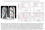
Der Beitrag stellt die mathematische Rekonstruktion der volumetrischen Wärmekarte und einer geeigneten Pilotstudie an 13 ex vivo Bioproteinphantomen mit flexiblen Schläuchen detailliert vor. Die flexiblen Schläuche simulieren dabei einen Kühlkörpereffekt, der in vivo durch die Gewebe-durchziehenden Blutgefäße besteht und sich auf die Hitzeverteilung während der Thermoablation auswirkt. Die Auswertung zeigt vielversprechende Ergebnisse in Bezug auf die Ähnlichkeit der Rekonstruktion im Vergleich zur realen Koagulationsnekrose. Zum Vergleich zeigt die Abbildung links die Wärmekarte mit einem Wärmegradienten von rot (warm) nach grün (kalt), in der Mitte die aus dieser Wärmekarte berechnete Zone des zerstörten Gewebes und rechts die medizinisch ermittelte, reale Zone des zerstörten Gewebes (Koagulationsnekrose).
Detaillierte Vorab-Infos sind hier zu finden.
(September 2021)
--------------------------------------
2.5D thermometry imaging for MR-guided tumor ablation @ MICCAI 2021
For this year's MICCAI conference from September 27th until October 1st, the team of researchers with Julian Alpers and Daniel Reimert will present their latest results on image-guided tumor ablation using 2.5D thermometry imaging in the MRI. With an impact factor of 8.5, this conference is one of the most important and far-reaching international conferences in the field of "Medical Image Processing and Computer-Aided Intervention". Overall, the MICCAI Society is dedicated to the promoting, sustaining, and facilitating of research, education and practice in the fields of medical image processing and computational medical interventions including biomedical imaging and medical robotics.
The contribution to be presented from the STIMULATE research campus deals with the urgent clinical need for fast and reliable monitoring of the volumetric heat distribution during MRI-guided tumor ablation. The researchers were able to develop a method for generating 2.5D thermometric maps. A so-called 2.5D image is created from evenly distributed 2D MRT phase images rotated around the applicator’s main axis. The images can be fetched directly from the MR device, which reduces the delay between image acquisition and visualization. Medical staff benefit of the ablation assessment in real time, for example during tumor ablation. The aim of tumor ablation is a so-called A0 ablation, whereby as much diseased tissue as necessary and at the same time as little healthy tissue as possible is destroyed with a safety margin around the tumor.
The article presents the mathematical reconstruction of the volumetric heat map and a suitable pilot study on 13 ex vivo bioprotein phantoms with flexible tubes. The flexible tubes simulate a heat sink effect, which exists in vivo through the blood vessels permeating the tissue affecting the heat distribution during thermal ablation. The evaluation shows promising results with respect to the similarity of the reconstruction compared to real coagulation necrosis. In comparison, the figure on the left shows the heat map with a thermal gradient from red (warm) to green (cold), in the middle the zone the destroyed tissue calculated from this heat map and on the right the medically determined, real zone of destroyed tissue (coagulation necrosis).
Detailed information in advance can be obtained here.(September 2021)
 nach grün (kalt), Mitte: berechnete Zone des zerstörten Gewebes, rechts: medizinisch ermittelte, reale Zone des zerstörten Gewebes (Koagulationsnekrose)")
 Eröffnung